
![]() Views: 461
Views: 461
Ultrasound for Endometriosis: Procedure and Results

Social share:
However, it should be noted that ultrasound alone may not be able to confirm the diagnosis of endometriosis, and additional tests, such as laparoscopy, may be required.
Dedicated Support at Every Step!
Our Doctors are available 24 hours a day, 7 days a week to help you!
Table of Contents
How Does an Ultrasound Test Detect Endometriosis?
Ultrasound uses high-frequency sound waves to create images of the inside of the body. In the case of endometriosis, an ultrasound test may be used to detect the presence of abnormal tissue growth outside the uterus, which is a characteristic of the condition. A transvaginal ultrasound, which uses a wand-like device inserted into the vagina, is often used to get a better view of the pelvic area. The ultrasound can show the presence of endometrial tissue in areas where it shouldn't be, such as on the ovaries or fallopian tubes. However, it is worth noting that ultrasound is not always able to confirm a diagnosis of endometriosis and may be used in conjunction with other diagnostic procedures, such as laparoscopy.
No Cost EMI, Hassle-free Insurance Approval
What Does Endometriosis Look Like on Ultrasound?
Endometriosis can appear on ultrasound as small, dark lesions or "spots" on the ovaries, fallopian tubes, or other areas in the pelvic region. These lesions may also be described as "powder burn" or "powder burn-like" lesions. These lesions may appear as cystic masses on the ovary, and sometimes they can be difficult to differentiate from functional ovarian cysts.
On a transvaginal ultrasound, endometriomas (cysts caused by endometriosis) appear as dark, fluid-filled cysts with thick, irregular walls. They may also be described as having a "target" or "ring" appearance.
It's important to note that these results are not specific to endometriosis and can be caused by other conditions as well. Therefore, ultrasound should be used in conjunction with other diagnostic tools, such as laparoscopy.
4.5/5
24 Years Experience Overall
4.6/5
19 Years Experience Overall
4.6/5
19 Years Experience Overall
4.7/5
17 Years Experience Overall
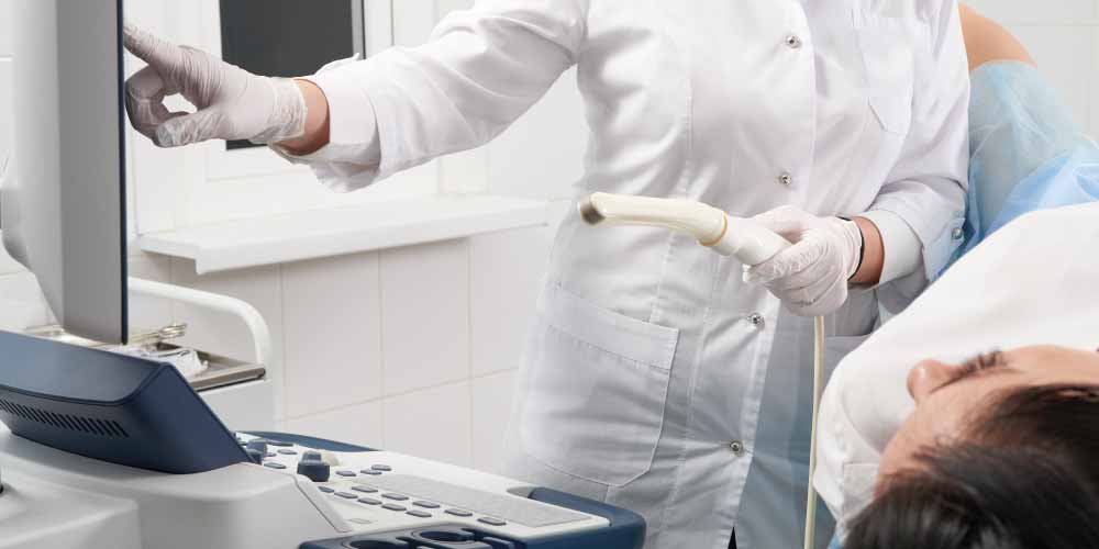
How is an Ultrasound Test Performed to Detect Endometriosis?
An ultrasound test to detect endometriosis is typically performed as a transvaginal ultrasound. This type of ultrasound uses a wand-like device (transducer) that is inserted into the vagina. The transducer sends out high-frequency sound waves that bounce off the internal organs and create images that can be viewed on a monitor.
During the test, the person will lie on their back with their feet in stirrups, and the transducer will be inserted into the vagina. The technologist will then use the wand to scan the pelvic area, including the uterus, ovaries, and fallopian tubes. The images will be viewed on a monitor in real-time, and the radiologist will take images of any areas of concern. The test typically takes 20-30 minutes.

It's important to note that the ultrasound test should be performed during a specific time of the menstrual cycle, as endometriosis can be hard to detect when it is not active. Therefore, it is best to perform the test in the middle of the menstrual cycle, around day 14-15, or during the luteal phase.
It's also important to note that the ultrasound test alone may not confirm the diagnosis of endometriosis, and other diagnostic tools, such as laparoscopy, may be needed.
Also Read: Which doctor to consult for endometriosis?
How Does the Doctor Predict the Result of an Ultrasound Test for Endometriosis?
Doctors use a combination of factors to predict the results of an ultrasound for endometriosis, including the patient's symptoms, physical examination findings, and the ultrasound images themselves.
- During an ultrasound, the doctor will look for characteristic signs of endometriosis, such as the presence of endometrial tissue outside of the uterus, cysts (endometriomas), and adhesions. The size, location, and number of these lesions can also provide clues about the severity of endometriosis.
- The doctor may also take into account the patient's symptoms, such as pelvic pain and painful periods, including her medical and family history of endometriosis.
Ultrasound is a useful diagnostic test for detecting endometriosis. If you are experiencing symptoms of endometriosis, then you must consult an experienced gynecologist. Visit Pristyn Care and consult our top gynecologists, who have ample experience in providing detailed diagnosis and treatment for endometriosis.
Author:


Dedicated Support at Every Step!
Our Doctors are available 24 hours a day, 7 days a week to help you!


