
![]() Views: 2,769
Views: 2,769
X-Ray to Diagnose Shoulder Dislocation
X-rays are electromagnetic radiations that provide an image of the body’s internal structures painlessly. Different tissues in the body absorb different amounts of radiation based on their composition. However, in general, the biggest difference can be seen between bones and the rest of the soft tissues.

Pristyn Care Team
Social share:
Dedicated Support at Every Step!
Our Doctors are available 24 hours a day, 7 days a week to help you!
Table of Contents
What is an X-Ray?
Bones generally appear white on X-ray because they absorb a lot of X-rays due to their high calcium concentration. On the other hand, fat, air, and other soft tissues absorb less amount of x-rays and look gray to black.
This apparent difference is the reason why x-rays are generally used to diagnose health issues related to the bones, such as fractures, joint dislocations, tumor formations, etc.
No Cost EMI, Hassle-free Insurance Approval
When is a Shoulder X-Ray performed?
A shoulder X-ray is performed to diagnose a health issue with the shoulder joint. It is completely non-invasive and normally minimal pain. However, if the patient has severe trauma, they might experience a little difficulty holding still for the x-ray.
Shoulder x-rays are performed for the following conditions:
- Shoulder dislocation
- Broken bones surrounding the shoulder joint
- Bone spurs near the shoulder joint
- Arthritic shoulder degeneration
- Benign or malignant tumors in the shoulder
- Bursitis
- Problems with the alignment of the shoulder joint
- Rotator cuff tear or calcifications
Who performs an X-Ray for Shoulder Dislocation?
X-rays are generally performed by a radiology technician or radiologist assistant (RA). Normally, after a shoulder dislocation, the patient should visit an orthopedic doctor, who will prescribe the required diagnostic or imaging tests, including X-ray. However, they can also visit a radiologist directly.
After the technician has completed the X-ray, the radiologist will look at the X-ray to make sure the image is not smudgy or has any other defects. The radiologist might inform you of the diagnosis themselves, or they may refer you to an orthopedist for further treatment.
Precautions to take before getting an X-Ray for Shoulder Dislocation
There are no dietary restrictions before a shoulder dislocation x-ray imaging test. However, metal objects like jewelry, body jewelry, glasses, hairpins, etc., can mess with the x-ray image, so they should be removed beforehand.
You might need to wear a protective lead collar or shield to protect sensitive body parts from harmful radiation. If you are pregnant, you should avoid getting X-rays. If it is unavoidable, then inform your radiologist so that they can prepare you accordingly.
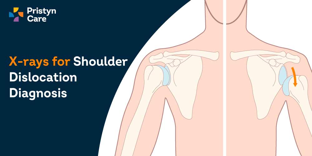
What is a Shoulder Dislocation?
Shoulder dislocation, also known as glenohumeral joint dislocation, occurs when the humerus separates from the glenoid of the scapula at the glenohumeral joint. Based on the direction of the dislocation, it can be classified as:
- Anterior shoulder dislocation (occurs in 95% of the cases)
- Posterior shoulder dislocation
- Inferior shoulder dislocation
All shoulder dislocations present with shoulder pain and reduced range of motion. They almost always occur due to a traumatic injury, like a sports injury, assault, seizure, or fall. They are managed via pain medication and immediate reduction.
How is a Shoulder X-Ray taken?
A shoulder x-ray usually takes less than 5 minutes and does not require any anesthetic. You may need to change into a hospital gown for the imaging. Based on the injury, your technician will direct you to either stand or sit while placing your shoulder next to the X-ray machine.
Your technician will then provide you with the appropriate lead covering to help you protect yourself from x-rays. You may have to hold still, hold your breath for short periods or move your shoulder into different positions during the imaging test.
If any of the images are blurry, then the technician will retake them. Digital x-rays are usually available straight away, but film x-rays can take 15-20 minutes to develop.
After the x-ray has been taken, you can change back into your clothes and put your jewelry and glasses back on.
How does Shoulder Sislocation appear on an X-Ray?
Shoulder dislocation and injury X-rays are performed in two views: Frontal and Y-views. In addition to the diagnosis of shoulder dislocation, the X-ray is also used to diagnose other injuries like fractures of the humeral head or neck or the clavicle. Generally, two x-rays views are taken for shoulder dislocation diagnosis:
Frontal view/AP view
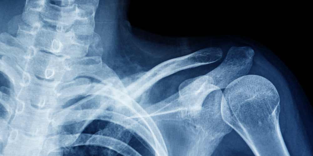
Normally, anterior and inferior shoulder dislocations can be easily diagnosed in a frontal view based on the position of the humerus head and the outline of the glenoid fossa. The humerus normally moves medially and overlies the glenoid in case of anterior dislocation.
Y-view
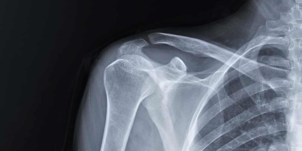
Posterior dislocations don’t normally appear on an AP (anteroposterior) view, as the humerus appears to maintain a proper position with the glenoid. As such, a trans-scapular Y view x-ray is needed. In a normal Y-view x-ray, the humerus appears centered over a Y-shape formed by the coracoid, blade of the scapula, and spine of the scapula (acromion).
What are the additional tests required for Shoulder Dislocation diagnosis?
In addition to normal radiographs, patients also need the following diagnostic tests for a complete diagnosis of shoulder dislocation:
- CT (computed tomography) scan for the assessment of associated complex fractures in the glenoid rim
- MRI (magnetic resonance imaging) to diagnose if the patient has shoulder instability due to a ligament or tendon injury in the labrum
- Point-of-care ultrasound, i.e., ultrasonography that is performed, interpreted and utilized simultaneously by the orthopedist during consultation for immediate diagnosis and management.
What are the diagnostic findings in a radiograph showing a Shoulder Dislocation?
Using an X-ray, the orthopedist can determine the following about a shoulder dislocation:
- Whether the patient has a dislocated joint
- The direction of the dislocation
- Fractures or other injuries associated with the dislocation
- Hill-Sachs defect, i.e., fracture of the humerus
- Bony Bankart lesion, i.e., detachment of the anteroinferior labrum along with a glenoid rim fracture
- Acromioclavicular joint disruption (dislocation)
- Acromial fracture
- Proximal humerus fracture
- Clavicular fracture
In addition to these, the ribs and a part of the lungs are also included in a shoulder x-ray and can help find other issues such as pneumothorax, injury to soft tissue structures like the neurovascular bundle, etc.
What are the risks associated with a Shoulder X-Ray?
Normally, x-rays expose you to a very small amount of radiation. Therefore, there is very little risk of them causing tissue damage or cancerous mutation. Though x-rays are normally safe, some areas, like the thyroid glands, testes, breast tissue, etc., are more susceptible to radiation damage.
Additionally, children and developing fetuses are more sensitive to X-ray radiation. For children, radiologists generally use lower dosages of radiation while taking an x-ray. But if you are pregnant, you should discuss the advantages and disadvantages of the test with your doctor before getting an X-ray.
Normally, radiology technicians provide the patient with a lead apparatus that they can use to protect their sensitive areas. You should always ensure you are wearing the appropriate padding while taking an x-ray

Dedicated Support at Every Step!
Our Doctors are available 24 hours a day, 7 days a week to help you!


