Select City

Learn about retinal detachment surgery, treatment options, recovery, cost, insurance coverage, retinopathy of prematurity, and when to consult a retina specialist for urgent care.
Learn about retinal detachment surgery, treatment options, recovery, cost, insurance coverage, retinopathy of ... Read More


Free Cab Facility

No-Cost EMI

Support in Insurance Claim

1-day Hospitalization

USFDA-Approved Procedure
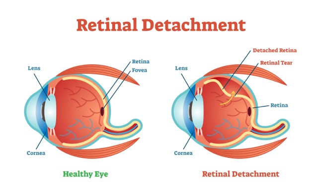
Retinal detachment is a serious eye condition that occurs when the retina the light sensitive layer at the back of the eye pulls away from its normal position. Without prompt treatment, retinal detachment can lead to permanent vision loss. Fortunately, modern retinal detachment surgery can successfully reattach the retina and preserve vision in many patients. Early diagnosis by an experienced retina specialist is critical for achieving the best outcomes.
Disease name
Surgery name
Duration
Treated by
Free Retinal Detachment Surgery Cost Calculator
Fill details to get actual cost
The retina converts light into signals that are sent to the brain, allowing us to see. When the retina detaches from the underlying tissue, its blood and oxygen supply becomes compromised.
Retinal detachment is considered an ophthalmic emergency and requires immediate medical attention.
Rhegmatogenous Retinal Detachment
This is the most common type and occurs when a retinal tear or hole allows fluid to collect beneath the retina.
Tractional Retinal Detachment
This occurs when scar tissue on the retinal surface pulls the retina away from the back of the eye. It is commonly seen in advanced diabetic eye disease.
Exudative Retinal Detachment
Fluid accumulates beneath the retina without a tear, often due to inflammation, tumors, or vascular disorders.
Recognizing symptoms early can help prevent permanent vision loss.
Any sudden visual changes should be evaluated by a retinal detachment doctor immediately.
Diet & Lifestyle Consultation
Post-Surgery Recovery Follow up
Free Cab Facility

24*7 Patient Support
Several factors can increase the risk of retinal detachment.
Common Causes
Retinal detachment requires urgent evaluation by a retina specialist.
You should seek immediate medical attention if you experience:
Prompt intervention can significantly improve the chances of preserving vision.
A comprehensive eye examination is required to confirm retinal detachment.
Special eye drops enlarge the pupil, allowing the ophthalmologist to examine the retina thoroughly.
OCT creates detailed cross-sectional images of retinal layers.
Ultrasound imaging helps visualize the retina when bleeding or cataracts obstruct direct examination.
High resolution retinal images help document retinal abnormalities.
The treatment approach depends on the severity and type of retinal detachment.
Laser Photocoagulation
Laser treatment seals small retinal tears before detachment develops.
Cryopexy
A freezing treatment used to seal retinal tears and prevent progression.
Pneumatic Retinopexy
A gas bubble is injected into the eye to push the retina back into place while the retinal tear is sealed.
Scleral Buckling Surgery
A silicone band is placed around the eye to reduce traction and support retinal reattachment.
Vitrectomy Surgery
Vitrectomy is one of the most commonly performed retinal detachment surgeries. The surgeon removes the vitreous gel, repairs the retina, and uses gas or silicone oil to support healing.
Before Surgery
Patients undergo:
During Surgery
The procedure may be performed under local or general anesthesia depending on the patient’s condition and surgical complexity.
After Surgery
Patients receive:
Recovery varies based on the severity of detachment and surgical technique.
Visual improvement may continue for several months. The final visual outcome depends on:
Many patients search for retinal detachment surgery cost before treatment.
The cost can vary depending on:
| Procedure | Estimated Cost Range |
|---|---|
| Laser Retinal Repair | ₹15,000 – ₹50,000 |
| Pneumatic Retinopexy | ₹30,000 – ₹80,000 |
| Scleral Buckling | ₹50,000 – ₹1,20,000 |
| Vitrectomy Surgery | ₹70,000 – ₹2,50,000+ |
Actual costs may vary depending on the patient’s condition and treatment plan.
Many health insurance policies cover retinal detachment treatment because it is considered medically necessary rather than cosmetic.
Coverage may include:
Patients should verify policy terms and pre-authorization requirements with their insurer before treatment.
Retinopathy of Prematurity (ROP) is a retinal disorder that affects premature infants.
Risk Factors for ROP
Why Early Screening Matters
Untreated ROP can lead to retinal detachment and permanent blindness. Early screening by pediatric retina specialists helps identify infants who require treatment.
The success of retinal detachment treatment often depends on timely intervention and specialist expertise.
Fellowship Training
Choose a retina specialist with advanced vitreoretinal surgery training.
Experience
Look for surgeons who regularly perform retinal detachment procedures.
Hospital Infrastructure
Advanced retinal imaging and surgical equipment can improve outcomes.
Emergency Availability
Retinal detachment is time-sensitive, making prompt access to care important.
Although retinal surgery is generally safe, potential risks include:
Regular follow up visits help detect and manage complications early.
Most patients can return to routine activities after recovery.
Tips for Long-Term Eye Health
The cost typically ranges from ₹50,000 to ₹2,50,000 or more, depending on the surgical procedure, hospital, and surgeon’s expertise.
Yes, most health insurance plans cover retinal detachment surgery when it is medically necessary.
Out of pocket expenses depend on policy coverage, deductibles, co payments, and hospital network status. Many patients may have a significant portion of the treatment covered.
A retinal detachment specialist is an ophthalmologist with advanced training in diseases and surgery of the retina and vitreous.
Claims may occasionally be denied due to policy exclusions, documentation issues, or lack of authorization. Patients can often appeal these decisions.
Common symptoms include flashes of light, floaters, blurred vision, peripheral vision loss, and a curtain-like shadow over vision.
Yes, recurrence is possible in some cases. Regular follow up appointments help detect and manage any new retinal problems early.

Sameer Khan

 Recommends
I had a retinal detachment scare and the emergency care at Healing Touch Hospital was outstanding.
trisha
Recommends
The Doctor is very experienced. Also staff is very helpful and cooperative.Thank you so much(pristyn care)for your support and guidance.
.svg)


.svg)